Research
Nucleation, growth and self-assembly are pervasive phenomena across chemistry, biology, and physics.
Overview
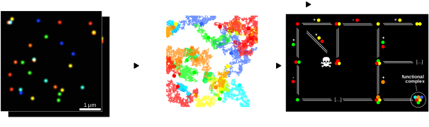
Many of these processes are currently technologically challenging to observe in both the biological and nanotechnological world, and thus remain poorly understood. Nanomaterial syntheses that could transform energy and medicine remain frustratingly empirical. The same protocol, run twice, gives different products: because nobody can watch the earliest, nanometre-scale moments where that variability is set. Transient assemblies of proteins, meanwhile, underpin everything from cell signalling to the immune response, but are too short-lived and too crowded with interacting partners for existing microscopes to resolve. We develop spectroscopic tools that make both newly observable.
Approach
Our research spans steady-state, time-resolved, and single-particle spectroscopies — with a key focus on the single-particle, since averaging over a population hides exactly the rare events and heterogeneity we want to understand. Development of these techniques encompasses two key aspects:
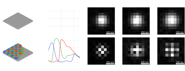
- Experimental — building optical tools such as photothermal microscopy (which images single nanometre-scale objects through their non-radiative decay, without any need for a fluorescent label) and multicolour single-molecule microscopy (which tracks several interacting molecular species at once by encoding their identity in the spectral domain).
- Analytical — applying statistics, information theory and chemical kinetics to turn those measurements into mechanism, extracting as much as possible from every photon we collect.
Nanoparticle nucleation and growth
Nanomaterials already underpin technologies in energy, catalysis and medicine, but their synthesis remains largely empirical — identical protocols can yield different products, because the earliest moments of particle formation (nucleation and the first steps of growth) have never been directly observed one particle at a time. We're developing photothermal microscopy sensitive enough to watch individual nanoparticles form and grow, starting with gold, to understand what actually controls the spread of outcomes a synthesis produces.
Transient biomolecule assembly
Short-lived, multi-partner protein interactions drive processes like cell signalling and immune activation, but existing microscopy can't follow more than one or two interacting species at once in their native environment. We're building spatial-spectral single-molecule microscopy — which separates many molecular species by colour in a single image — to watch these assemblies form in real time. We are currently collaborating on doing so in respiratory supercomplexes.